Support your step, relieve flatfoot
Adult Flatfoot
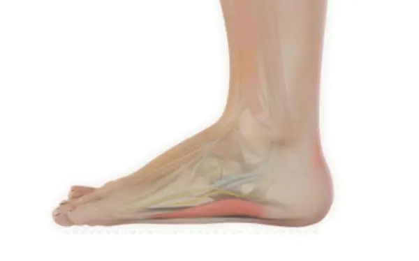
Posterior tibial tendon dysfunction is one of several terms to describe a painful, progressive flatfoot deformity in adults. Other terms include posterior tibial tendon insufficiency and adult acquired flatfoot.
The term adult acquired flatfoot is more appropriate because it allows a broader recognition of causative factors, not only limited to the posterior tibial tendon, an event where the posterior tibial tendon loses strength and function.
The adult acquired flatfoot is a progressive, symptomatic (painful) deformity resulting from gradual stretch (attenuation) of the tibialis posterior tendon as well as the ligaments that support the arch of the foot.
What Causes Adult Flatfoot?
Adult acquired flatfoot, also known as posterior tibial tendon dysfunction (PTTD), develops when the posterior tibial tendon — the primary tendon supporting the foot’s arch — gradually weakens or tears. This condition is especially common in middle-aged and older adults, particularly those who are overweight or have poor circulation due to aging or atherosclerosis.
Over time, the weakened tendon can no longer support the arch, causing the surrounding ligaments to stretch and tear. The bones in the arch shift out of position under body weight, leading to pronation — an inward rolling of the foot and ankle. As the deformity progresses, the arch collapses, and the heel bone tilts inward, potentially causing the foot to dislocate outward from under the ankle joint.
Contributing Risk Factors Include:
- Age-related weakening of tendons and ligaments
- Obesity or excess weight
- Poor circulation (arterial narrowing)
- Biomechanical stress over time
- Repetitive strain on the posterior tibial tendon
Stages of Adult Acquired Flatfoot
At SoCal Foot and Ankle Doctors, with offices in Santa Monica and the Cedars-Sinai Medical Towers, we classify adult acquired flatfoot into three stages:
- Stage I: Inflammation and swelling of the posterior tibial tendon along the inside of the ankle. Pain may be present, but the foot remains flexible.
- Stage II: Visible flattening and deformity of one foot compared to the other. The arch collapses, but the deformity is still flexible and correctable.
- Stage III: The flatfoot deformity becomes rigid and painful, especially along the outside of the ankle, often limiting mobility.
How Is Adult Flatfoot Diagnosed?
A precise diagnosis begins with a clinical examination by a podiatrist experienced in flatfoot disorders. At our Santa Monica and Cedars-Sinai locations, we assess:
- Gait and posture (how the patient walks)
- Muscle strength testing
- Foot deformity comparison (symptomatic vs. unaffected foot)
Common Office Test: Single Heel Raise
The patient stands on the affected foot and attempts to rise onto their toes. In advanced PTTD, the heel fails to lift or does not invert — a key sign of posterior tibial tendon injury.
Imaging:
- X-rays: Useful for assessing bone alignment, but not definitive for tendon injury.
- MRI: Can visualize tendon damage or inflammation but is not always necessary.
- Ultrasound: Sometimes used to detect tendon changes, though less commonly.
Treatment for Adult Acquired Flatfoot
Early treatment is critical to prevent long-term deformity and disability. At SoCal Foot and Ankle Doctors, we focus on non-surgical treatments whenever possible:
Stage I – Conservative Care:
- Rest and activity modification
- Short-term casting or walking boot
- Anti-inflammatory therapy
- Custom-molded orthotics and supportive footwear
Stage II – Supportive Bracing:
- Continued use of orthotics
- Ankle-foot orthosis (AFO), such as the Richie Brace
- This custom brace is lightweight, fits easily into athletic footwear, and provides arch support and ankle stability.
Stage III – Surgical Intervention (if conservative care fails):
Surgery may be recommended to:
- Alleviate pain
- Halt deformity progression
- Restore foot alignment and function
Procedures include:
- Tendon debridement or transfer
- Osteotomy (bone realignment)
- Joint fusion (arthrodesis) in advanced cases
Recovery often requires 6–12 weeks in a cast and non-weight bearing for up to 8 weeks after fusion procedures.
When to See a Specialist
If you’re experiencing arch pain, ankle weakness, or your foot is flattening over time, schedule a consultation at SoCal Foot and Ankle Doctors. We serve patients from Santa Monica, Los Angeles, and those seeking trusted care near Cedars-Sinai Medical Center.
Don’t wait until surgery is your only option. With early intervention — including custom orthotics, bracing, and targeted therapies — many patients can avoid surgery and restore foot function successfully.
Our Docters
Our Board Certified Podiatrists
Socal Foot and Ankle doctors are committed to delivering the most exceptional treatments.

Board Certified Foot & Ankle Specialist
Office Time
Location: Santa Monica
Mon – Thur: 9:00 AM – 5:00 PM
Friday: 9:00 AM – 5:00 PM

Board Certified Foot & Ankle Specialist
Office Time
Location: Santa Monica
Mon – Thur: 9:00 AM – 5:00 PM
Friday: 9:00 AM – 5:00 PM
Request Appointment
NON-INVASIVE ADVANCED TREATMENT
BOARD CERTIFIED
FOOT & ANKLE
Surgeons
Years Experience
Happy Patients
Location
Local Partners
Request Appointment
NON-INVASIVE ADVANCED TREATMENT
BOARD CERTIFIED
FOOT & ANKLE
Surgeons
Years Experience
Happy Patients
Location
Local Partners