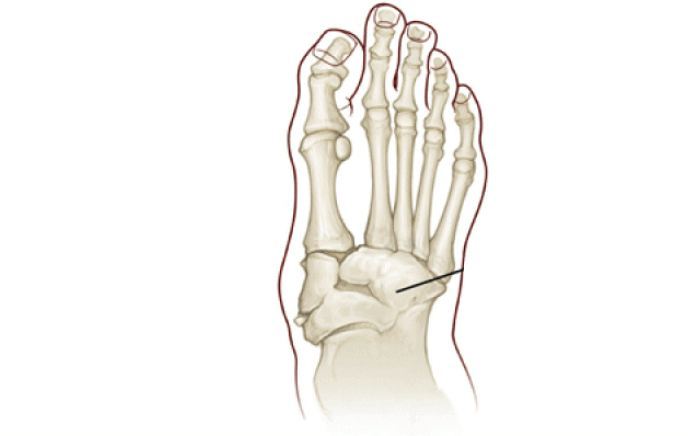
Progressive Degeneration Of A weight Bearing Joint
NEUROPATHIC ARTHROPATHY
Charcot joint disease was given its name by the French neurologist Jean-Marie Charcot in 1868. He noted a bizarre pattern of bone destruction in patients with tertiary syphilis and absent sensation. In 1936, William Jordan described a similar pattern in patients with diabetes.
Theories as to the cause of the Charcot joint disease abound. However, certain predisposing factors appear to be necessary in order for Charcot to develop. First, peripheral sensory neuropathy or total absence of sensation must be present. Second, circulation is most commonly normal. Third, there is often a history of a preceding injury. This is often so minimal the patient is unable to recall any injury or trauma.

Who Gets Charcot Disease?
Charcot joint disease was originally described in patients with tertiary syphilis and absent sensation. However, once penicillin was discovered, the incidence of syphilis dropped dramatically. In general, any disease process that results in loss of sensation in the lower extremity can lead to Charcot joint disease. Today, the leading cause of Charcot joint disease is diabetes mellitus. It is estimated that 1 out of 700 patients with diabetes will develop Charcot joint. Other entities, which can lead to Charcot joint, include chronic alcoholism, leprosy, hereditary insensitivity to pain, syringomyelia and multiple sclerosis.
A second key component to the development of Charcot is the history of a preceding injury to trauma to the foot or ankle. Often this injury goes undetected because of a lack of normal sensation. Additionally, because of poor sensation, the injury may not be perceived to be serious. Consequently, the patient will continue with normal or near-normal activity leading to further fracture and dislocation of the involved joints and bones. Patients who develop Charcot joint will also have good to excellent circulation to their feet. It is uncommon for patients with significant peripheral vascular disease to develop Charcot joint disease.
What Are The Symptoms Of Charcot Disease?
Patients who develop Charcot joint disease most commonly notice unexplained swelling in their foot or ankle. Because of the underlying loss of sensation, this swelling is often painless. On occasion, there may be redness localized to the top of the foot or ankle. There will also be increased warmth to the foot, indicating localized inflammation. Rarely will there be bruising unless the injury is significant.Because Charcot joint disease shows swelling and redness, it is often misdiagnosed. The most common misdiagnoses include cellulitis, osteomyelitis, tendonitis, and gout. Failure to diagnosis and treat this entity early and appropriately can lead to foot and ankle deformity
Diagnosing Charcot Joint Disease
The diagnosis is primarily based on the clinical examination. A diagnosis of Charcot joint must be considered in patients with diabetes who present with a warm, swollen foot or ankle without pain. Another critical feature in the diagnosis of Charcot joint is the presence of crepitus or “grinding” in the involved joints. This represents the unstable bone fragments moving against each other.
X-rays at this stage will often confirm the diagnosis, as these will often show fragmentation of bone and disruption of joints. Occasionally, if the x-rays fail to show any disruption and a diagnosis of Charcot joint disease is still being considered, bone scans can be helpful.
Special studies such as CT scans or MRI are rarely necessary to make the diagnosis. A CT scan can be useful if reconstructive surgery is planned. An MRI can be helpful if an infection with abscess formation is suspected.
Charcot Disease Treatment
Early diagnosis and treatment are critical to a successful outcome. Once the diagnosis of Charcot joint disease is made, initial treatment should consist of total non-weightbearing and immobilization of the involved extremity. This often requires the use of crutches or a walker. Additionally, a removable cast or brace to protect and immobilize the foot and ankle may be necessary. The duration of non-weightbearing and immobilization will depend on the joints affected and the degree of destruction. As a rule, the larger the joint, the longer the duration of non-weightbearing needs to be.
Serial x-rays and the resolution of the clinical features of the disease determine the healing of the involved joints. Clinically, one would expect to see a reduction of swelling, a decrease in skin temperature and a decrease in joint crepitus. On x-rays, one would expect to see resorption of bone debris and fragments with deposition of new bone and fracture healing. Protected weightbearing may be initiated when the clinical features of the disease show improvement, especially loss of joint crepitus. Weightbearing can be increased gradually as long as there is no re-exacerbation of the disease.When foot and ankle deformity develops, custom orthoses and special shoes may be necessary to prevent foot ulcerations and provide stability during ambulation. When ulcerations develop and resist conservative treatment, surgery may be necessary to prevent loss of the foot. Additionally, if there is severe instability, reconstructive surgery of the foot and ankle may be necessary to provide a stable platform for ambulation and to avoid lower leg amputation
Complications
The most common complication of Charcot joint disease is foot and ankle deformity. This can occur even following early and appropriate treatment. This typically results from significant bone and joint destruction that the functional integrity of the foot is sacrificed. Chronic ulcerations may occur as a result of these deformities. Surgery may be necessary to prevent limb loss. Another complication that may occur is the instability of the foot and ankle during ambulation. This once again is related to the joints affected and the degree of bone destruction. Severe instability can lead to lower leg amputation. Major reconstructive surgery may be necessary to prevent this complication of Charcot joint disease.
Our Board Certified Podiatrists
Socal Foot and Ankle doctors are committed to delivering the most exceptional treatments.

Board Certified Foot & Ankle Specialist
Office Time
Location: Santa Monica
Mon – Thur: 9:00 AM – 5:00 PM
Friday: 9:00 AM – 5:00 PM
Location Marina Del Rey
Mon – Thur: 9:00 AM – 5:00 PM
Friday: 9:00 AM – 5:00 PM
Location: Cedars Sinai
Mon – Thur: 9:00 AM – 5:00 PM
Friday: 9:00 AM – 5:00 PM

Board Certified Foot & Ankle Specialist
Office Time
Location: Santa Monica
Mon – Thur: 9:00 AM – 5:00 PM
Friday: 9:00 AM – 5:00 PM

BOARD CERTIFIED
FOOT & ANKLE
Surgeons
- Comprehensive Treatment of Foot & Ankle Conditions in the Pediatric, Adult & Geriatric population
- 3 Practice Locations Santa Monica Medical Plaza, Cedars Sinai Medical Towers, & UCLA Health in Marina Del Rey
- On Staff with Providence Saint Johns Health Center &Cedars Sinai Medical Center
