HEEL SPUR
What is Heel Spur Syndrome?
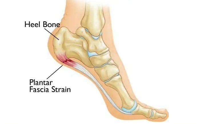
A heel spur is a pointed bony outgrowth of the bone of the heel (the calcaneus bone). Chronic local inflammation at the insertion of soft-tissue tendons or plantar fascia is a common cause of bone spurs (osteophytes). Heel spurs can be located at the back of the heel or under the heel, beneath the arch of the foot. Heel spurs at the back of the heel are frequently associated with inflammation of the Achilles tendon (tendinitis) and cause tenderness and heel pain made worse while pushing off the ball of the foot.
What Causes Heel Spur Or Heel Pain?
To understand the cause of the pain, one must understand the anatomy of the foot and some basic mechanics in the function of the foot. A thick ligament, called the plantar fascia, is attached from the bottom of the heel, fanning out into the ball of the foot, and attaching into the base of the toes. The plantar fascia is made of dense, fibrous connective tissue that will stretch very little. It acts as something like a shock absorber. As the foot impacts the ground with each step, the foot flattens out, lengthening the foot. This action pulls on the plantar fascia, which stretches slightly. When the heel comes off the ground, the tension on the ligament is released. Anything that causes the foot to flatten excessively will cause the plantar fascia to stretch greater than it is accustomed to. One consequence is the development of small tears where the ligament attaches into the heel bone. When these small tears occur, a very small amount of bleeding occurs and the tension of the plantar fascia on the heel bone produces a spur on the bottom of the heel. Pain experienced in the bottom of the heel is not produced by the presence of the spur. The pain is due to excessive tension of the plantar fascia as it tears from its attachment into the heel bone. Heel spur formation is secondary to the excessive pull of the plantar fascia where it attaches to the heel bone. Many people have heel spurs at the attachment of the plantar fascia with out having any symptoms or pain. There are some other relatively uncommon causes of heel pain.
There are several factors that cause the foot to flatten and excessively stretch the plantar fascia. The primary factor is the structure of a joint complex below the ankle joint, called the subtalar joint. The movement of this joint complex causes the arch of the foot to flatten and to heighten. Flattening of the arch of the foot is termed pronation and the heightening of the arch is called supination. If there is excessive pronation of the foot during walking and standing, the plantar fascia is strained. Over time, this will cause a weakening of the ligament where it attaches into the heel bone and causes pain. When a person is at rest and off of their feet, the plantar fascia attempts to mend itself. Then with the first few steps, the fascia re-tears causing pain. Generally, after the first few steps, the pain diminishes. This is why the heel pain tends to be worse the first few steps in the morning or after rest.
Another factor that contributes to the flattening of the arch of the foot is the tightness of the calf muscles. The calf muscle attaches into the foot by the Achilles tendon into the back of the heel. When the calf muscle is tight, it limits the movement of the ankle joint. When ankle joint motion is limited by the tightness of the calf muscle, it forces the subtalar joint to pronate excessively. Excessive subtalar joint pronation can cause several different problems to occur in the foot. In this instance, it results in excessive tension of the plantar fascia. The tightness of the calf muscles can be a result of several different factors. Exercise, such as walking or jogging, will cause the calf muscle to tighten. Inactivity or prolonged rest will also cause the calf muscle to tighten. Women who wear high heels and men who wear western style cowboy boots will, over time, develop tightness in the calf muscles.
Heel Spur Diagnosis
The diagnosis of heel pain and heel spurs is made by a thorough history of the course of the condition and by physical exam. Weight-bearing x-rays are useful in determining if a heel spur is present and to rule out rare causes of heel pain such as a stress fracture of the heel bone, the presence of bone tumors or evidence of soft tissue damage caused by certain connective tissue disorders.
Heel Spur Treatment
Treatments for heel spurs may include:
- Rest: Getting plenty of rest and taking pressure off the feet can help to reduce pain and swelling in the affected area.
- Applying ice: This can help reduce pain and swelling.
- Using custom-made orthotics (shoe inserts): These donut-shaped inserts go inside the shoe to take the pressure off the heel.
- Wearing cushioned sports shoes: These may also help to relieve pressure and reduce pain.
- Anti-inflammatory medication: This helps to reduce swelling.
- Cortisone injections: These reduce swelling and pain in the affected area. They are a stronger option if over-the-counter anti-inflammatory medication is not effective.
- In rare cases, surgery may be necessary to remove the heel spur. However, the above treatments are usually effective, and surgery is not needed.
If heel spurs are caused by an inflammatory type of arthritis, treatments for the underlying condition may also improve symptoms.
Adult Flatfoot Treatment
The adult acquired flatfoot is best treated early. There is no recommended home treatment other than the general avoidance of prolonged weight-bearing in non-supportive footwear until the patient can be seen in the office of the foot and ankle specialist.
In Stage I, the inflammation and tendon injury will respond to rest, protected ambulation in a cast, as well as anti-inflammatory therapy. Follow-up treatment with custom-molded foot orthoses and properly designed athletic or orthopedic footwear are critical to maintaining the stability of the foot and ankle after initial symptoms have been calmed.
Once the tendon has been stretched, the foot will become deformed and visibly rolled into a pronated position at the ankle. Non-surgical treatment has a significantly lower chance of success. Total immobilization in a cast or Camwalker may calm down symptoms and arrest the progression of the deformity in a smaller percentage of patients. Usually, long-term use of a brace known as an ankle-foot orthosis is required to stop the progression of the deformity without surgery.
A new ankle foot orthosis is known as the Richie Brace is available from a number of podiatric labs (www.Richiebrace.com), has proven to show significant success in treating Stage II posterior tibial dysfunction and the adult acquired flatfoot. This is a sport-style brace connected to a custom corrected foot orthotic device that fits well into most forms of lace-up footwear, including athletic shoes. The brace is lightweight and far more cosmetically appealing than the traditional ankle foot orthosis previously prescribed.
In cases where cast immobilization, orthoses, and shoe therapy have failed, surgery is the next alternative. The goal of surgery and non-surgical treatment is to eliminate pain, stop the progression of the deformity and improve mobility of the patient. Opinions vary as to the best surgical treatment for adult acquired flatfoot. Procedures commonly used to correct the condition include tendon debridement, tendon transfers, osteotomies (cutting and repositioning of bone) and joint fusions. (See surgical correction of adult acquired flatfoot)
Patients with adult acquired flatfoot are advised to discuss thoroughly the benefits vs. risks of all surgical options. Most procedures have long-term recovery mandating that the correct procedure be utilized to give the best long-term benefit. Most flatfoot surgical procedures require six to twelve weeks of cast immobilization. Joint fusion procedures require eight weeks of non-weight bearing on the operated foot – meaning you will be on crutches for two months.
The bottom line is: Make sure all of your non-surgical options have been covered before considering surgery. Your primary goals with any treatment are to eliminate pain and improve mobility. In many cases, with the properly designed foot orthosis or ankle brace, these goals can be achieved without surgical intervention.
Our Docters
Our Board Certified Podiatrists
Socal Foot and Ankle doctors are committed to delivering the most exceptional treatments.
Board Certified Foot & Ankle Specialist
Office Time
Location: Santa Monica
Mon – Thur: 9:00 AM – 5:00 PM
Friday: 9:00 AM – 5:00 PM
Board Certified Foot & Ankle Specialist
Office Time
Location: Santa Monica
Mon – Thur: 9:00 AM – 5:00 PM
Friday: 9:00 AM – 5:00 PM
Request Appointment
NON-INVASIVE ADVANCED TREATMENT
BOARD CERTIFIED
FOOT & ANKLE
Surgeons
Years Experience
Happy Patients
Location
Local Partners